CT stands for ‘computed tomography’. Tomography is a medical imaging technique in which multiple x-ray images are taken in thin cross sections along a person’s body from a number of different angles. These 2 dimensional x-ray images can then be computerised to produce a 3-dimensional view of patients’ body.
CT scans are common. They show the bony structures in greater detail than plain x-rays. However, they do not show the nerves and discs in as great detail as MRI.
How is it done?
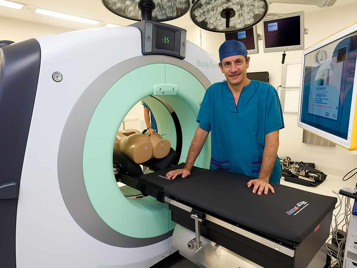
The test is performed by a radiographer (x-ray technician). The scanner obtains images using an x-ray beam which moves in a circle around the x-ray table taking multiple images in quick succession, while the table moves within the CT tunnel. The x-ray images are sent to a computer, operated by the radiographer, which processes the series of pictures, creating cross-sectional images or slices of the body. These images are displayed on a monitor and reviewed by a radiologist, (a doctor who is an x-ray specialist) who then interprets and reports on the images.
What are the risks?
Like many types of radiology, a CT scan involves exposure to radiation. An increased lifetime cancer risk is a rare risk due to this exposure to x-rays. If you are pregnant or suspect you may be pregnant, you should speak with your doctor before proceeding with this type of scan.
What will happen during the test?
You will be asked to change into a gown and remove any jewellery, watches or other objects that may interfere with the scan. You will then be asked to lie on the scan table, which is moved into the CT machine tunnel. During the scan the bed will slide back and forwards within the tunnel. You will need to lie as still as possible. The scan only takes a few minutes for a single region of the body. When they are finished, the scans will be reported and then sent to your doctor.
